Playback speed
10 seconds
“FIRST" ARTHROSCOPIC LIGAMENTOPLASTY TECHNIQUE FOR SCAPHOLUNATE INSTABILITY
12,310 views
August 16, 2012
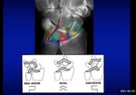
The treatment of scapholunate ligament injuries is complex and unreliable. Various open techniques have been ...
read more ↘ described for its treatment including capsulodesis, bone-ligament-bone transfers, tenodesis, partial fusions, proximal row carpectomies, etc(1). The development of wrist arthroscopy offers a new range of minimally invasive techniques including debridement, electrocoagulation and percutaneous fixation without opening the wrist(2,3). However, arthroscopic wrist ligament reconstruction has not been described before.
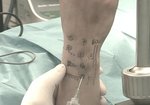
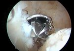
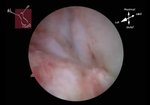
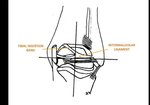
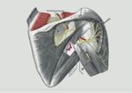
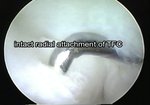
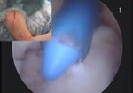
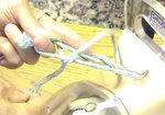
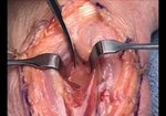
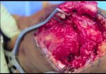
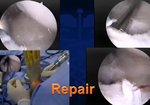
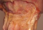
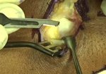
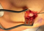
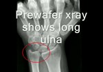
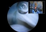
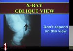
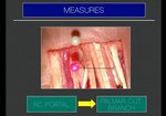
This video consists of four sections. The first one contains the description of the surgical technique for the arthroscopically reconstruction of the scapholunate ligament. The second one presents an anatomical study done in five cadaver specimens. The third one shows the technique performed in a patient. Finally, the last one includes the discussion and justification of the advantages and risks of the technique.
This technique tries to reproduce the “three-ligament tenodesis” in an arthroscopically manner(4). Two out of three objectives of the tenodesis are achieved: the reconstruction of the dorsal portion of the scapholunate ligament and the reinforcement of the volar and distal capsules. However, this technique does not tighten the dorsal radiotriquetral ligament. Therefore, its injury is a contra-indication for this technique.
The anatomical study performed demonstrates that the soft tissue injury is minimal compared with open reconstructions: 17 mm dorsal approach, 14 mm extensor retinaculum opening and 6 mm capsulotomy.
The posterior interosseous nerve was not damaged in any of the specimens.
There were no injuries to the radial artery or the superficial radial artery which were at an average of 15,6 mm and 10,6 mm respectively from the exit of the tunnel in the scaphoid tubercle.
The ligament reconstruction was always located at the dorsal portion of the scapholunate ligament, with mean thickness of 4 mm and mean length of 20 mm.
The indications for this arthroscopic ligamentoplasty are very clear: Geissler lesions(5) type III and IV showing dynamic or static instability that is easily reducible percutaneously.
With this arthroscopic technique we have three main objectives aimed at improving current open techniques:
- Reduce soft tissue injury by minimising dissection and thus greater postoperative mobility should be achieved.
- Avoid posterior interosseous nerve injury, thus maximising the proprioception of the wrist and the role of the dynamic stabilizers.
- Make an arthroscopic tenodesis that ensures optimal placement, tension and functionality of the ligament reconstruction.
We are confident that our proposal is technically feasible in a systematic and reproducible manner, with wide margin of safety.
1. Kuo, C. E. and S. W. Wolfe (2008). Scapholunate instability: current concepts in diagnosis and management J Hand Surg Am 33(6): 998-1013.
2. Shih, J. T. and H. M. Lee (2006). Monopolar radiofrequency electrothermal shrinkage of the scapholunate ligament. Arthroscopy 22(5): 553-557.
3. Mathoulin, C. and J. Messina (2010). Treatment of acute scapholunate ligament tears with simple wiring and arthroscopic assistance Chir Main 29(2): 72-77.
4. Garcia-Elias, M., A. L. Lluch, et al. (2006). Three-ligament tenodesis for the treatment of scapholunate dissociation: indications and surgical technique. J Hand Surg Am 31(1): 125-134
5. Geissler, W. B. (2006). Arthroscopic management of scapholunate instability. Chir Main 25 Suppl 1: S187-196.
↖ read less
Comments 23
Login to view comments.
Click here to Login

