Video Player is loading.
Current Time 0:00
/
Duration 0:00
Loaded: 0%
Stream Type LIVE
Remaining Time -0:00
1x
- 0.5x
- 0.75x
- 1x, selected
- 1.25x
- 1.5x
- 1.75x
- 2x
- Chapters
- descriptions off, selected
- captions settings, opens captions settings dialog
- captions off, selected
This is a modal window.
Beginning of dialog window. Escape will cancel and close the window.
End of dialog window.
10 seconds
Playback speed
This is a modal window. This modal can be closed by pressing the Escape key or activating the close button.
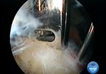
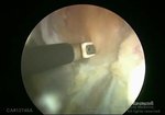
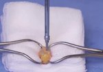
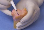
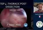
The Arthroscopically Assisted Latarjet Procedure in the Lateral Decubitus Position
1,906 views
May 14, 2013
Arthroscopic modifications of Latarjet procedure are becoming increasingly popular. We describe an arthroscopically assisted technique ...
read more ↘ in lateral decubitus position of the patient, which is still widely used by surgeons .
We try to develop a surgical technique, which allows to perform Latarjet procedure arthroscopically in lateral decubitus position of the patient through 3 standard portals, to save rotator interval, glenohumeral ligaments, joint labrum and capsule and to refixe them after coracoid transfer to the anterior – inferior defect of glenoid.
27 patients with anterior – inferior instability and massive deficit of glenoid were treated with our technique. After artroscopic evaluation and refreshing of glenoid cavity coracoid was osteotomized with the conjoined tendon and pulled out through the 2,5-3 cm incision. With the scope, pulled in the incision, the brachial plexus was visualized and palpated. The coracoid was passed through the supscapularis and fixed to the glenoid with 2 cannulated screws. Joint capsule and labrum were reattached to the glenoid rim and fixed with anchors.
The post-operative results were evaluated clinically, by X-ray and with 3D reconstruction. There was no recurrence of instability at the most recent follow-up (mean,18 months).
This technique allows to simplify arthroscopic Latarjet technique and to perform it in the lateral decubitus position. It decreases the possibility of subscapularis and axillary nerve damage, because of the palpation and visualization through the graft harvest incision. This technique also allows to save rotator interval , inferior and middle glenohumeral ligaments and to fixe joint capsule and labrum.
↖ read less
Comments 7
Login to view comments.
Click here to Login