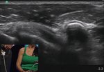
Scan Technique for Your Distal Biceps Tendon MSK Ultrasound Exam
By
Learn MSK Sono
FEATURING
Jamie Bie, RMSKS, RVT, RDMS
By
Learn MSK Sono
FEATURING
Jamie Bie, RMSKS, RVT, RDMS
80 views
July 24, 2023
The distal biceps tendon is one of the most difficult tendons in the body to ...
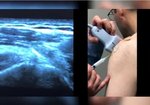
read more ↘ scan due to the oblique course of the tendon. This obliquity causes a lot of anisotropy during the ultrasound scan. For this reason, it is important to know the course of the tendon prior to starting your exam. You can actually use the anisotropy to your favor when mapping out the course of the tendon. The hypoechogenicity of the tendon makes it easier to visualize among the surrounding structures, but should be corrected before storing any images.
Here are my technical tips for improved visualization of the distal biceps tendon:
🔹Flex the patient’s arm.
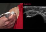
🔹Scan from the ulnar aspect of the arm, but angle the transducer radially towards the distal biceps tendon.
🔹Use the brachial artery as a window to visualize the distal biceps tendon. Fluid is advantageous for visualization during an ultrasound exam.
🔹In the long axis, heel the probe, and in the short axis toggle the probe upwards. Apply additional pressure while using these techniques.
Watch my scanning demonstration to see how it’s done!
↖ read less
Comments 0
Login to view comments.
Click here to Login


