Video Player is loading.
Current Time 0:00
/
Duration 0:00
Loaded: 0%
Stream Type LIVE
Remaining Time -0:00
1x
- 0.5x
- 0.75x
- 1x, selected
- 1.25x
- 1.5x
- 1.75x
- 2x
- Chapters
- descriptions off, selected
- captions settings, opens captions settings dialog
- captions off, selected
This is a modal window.
Beginning of dialog window. Escape will cancel and close the window.
End of dialog window.
10 seconds
Playback speed
This is a modal window. This modal can be closed by pressing the Escape key or activating the close button.
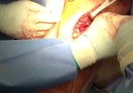
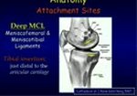
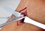
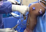
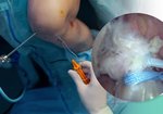
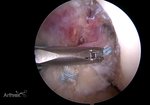
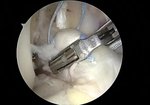
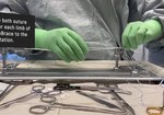
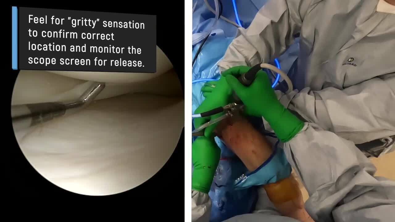
Quick Tips: Pie-Crusting the MCL to Improve Access to the Medial Knee Compartment
By
PEARL in Sports Medicine
FEATURING
Nathan Skelley
By
PEARL in Sports Medicine
FEATURING
Nathan Skelley
7,528 views
October 12, 2023
Dr. Skelley demonstrates a method for improving access to the medial compartment of the knee. ...
read more ↘ Important steps with this technique include being above the joint line and in the posterior medial region of the femur. The arthroscopic camera light can assist with finding the correct location. With the leg in a valgus position to put the MCL on stretch, enter the skin at one location with the 18 gauge needle. Do not make multiple skin punctures. Through the one poke hole in the skin, feel for a "gritty" sensation while walking the needle anterior and posterior. The needle tip will periodically contact the bone and pie-crust the MCL in the process to improve medial compartment arthroscopic visualization. This technique has not led to any clinical MCL laxity or medial sided knee pain at final follow-up in our experience. Pie-crusting has many benefits beyond improved visualization. The technique also allows for protecting the medial compartment structures while using instruments and it facilitates performing various procedures as demonstrated in this video.
Thank you for your time and consideration of this video.
↖ read less
Comments 4
Login to view comments.
Click here to Login