Video Player is loading.
Current Time 0:00
/
Duration 0:00
Loaded: 0%
0:00
Stream Type LIVE
Remaining Time -0:00
1x
- 0.5x
- 0.75x
- 1x, selected
- 1.25x
- 1.5x
- 1.75x
- 2x
- Chapters
- descriptions off, selected
- captions settings, opens captions settings dialog
- captions off, selected
This is a modal window.
Beginning of dialog window. Escape will cancel and close the window.
End of dialog window.
10 seconds
Playback speed
This is a modal window. This modal can be closed by pressing the Escape key or activating the close button.
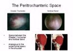
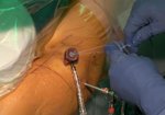
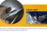
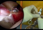
Peripheral Compartment First Hip Arthroscopy Technique
11,542 views
December 11, 2022
In this video, we discuss our technique for hip arthroscopy in which the peripheral compartment ...
read more ↘ is approached first. This is in contrast to starting the hip arthroscopy with the surgical leg on traction and using fluoroscopic images to access the central compartment with a needle as the first step in the hip arthroscopy procedure. We believe entering the peripheral compartment with a periportal technique improves visualization and access using the 30 degree scope. This approach is less likely to cause iatrogenic injury to the labrum and weight bearing cartilage in our experience. The technique mainly views from the DALA portal and works through the Dienst portal, however, the camera can be switched as needed to view and access the pathology with these portals. Special consideration needs to be given to portal entry point, capsular thinning, and leg/camera manipulation to view the necessary pathology. This approach also facilitates access to the central compartment which can be especially beneficial in cases with large pincer pathology. We demonstrate these techniques in the following video. Thank you for your time and consideration.
↖ read less
Comments 9
Login to view comments.
Click here to Login