Video Player is loading.
Current Time 0:00
/
Duration 0:00
Loaded: 0%
0:00
Stream Type LIVE
1x
- 0.5x
- 0.75x
- 1x, selected
- 1.25x
- 1.5x
- 1.75x
- 2x
- Chapters
- descriptions off, selected
- captions settings, opens captions settings dialog
- captions off, selected
This is a modal window.
Beginning of dialog window. Escape will cancel and close the window.
End of dialog window.
10 seconds
Playback speed
This is a modal window. This modal can be closed by pressing the Escape key or activating the close button.
2,559 views
August 17, 2011
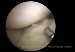
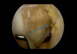
With the knee extended, plica was located between the patella and trochlea. At 30° of ...
read more ↘ flexion, the plica began to contact the medial femoral condyle, showing the bowstring phenomenon . At 60° of flexion, the plica began to slip from the medial femoral condyle, the so-called rolling-over sign. At 90° of flexion, the
plica slipped away from the medial femoral condyle. The MPP test was then repeated while maintaining
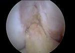
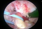
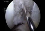
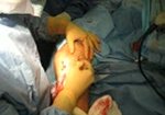
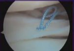
the superolateral viewing portal to confirm the phenomena of the test. When the affected knee was extended without digital compression, a fibrotic plica was seen apart from the medial femoral condyle. When manual force was applied to the inferomedial portion of the patellofemoral joint with the examiner’s thumb, the fibrotic plica was impinged between the patella and medial femoral condyle. Any other possible pathologic conditions can be assessed, including medial patellar or medial femoral condylar chondromalacia and synovitis.
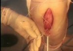
Excision of the thickened fibrotic portion of the pathologic plica was performed using a motorized shaver inserted through the anterolateral portal. Postoperative findings after excision of the MPP showed no patellotrochlear impingement without digital compression and with digital compression.
↖ read less
Comments 0
Login to view comments.
Click here to Login


