• 7 year old female with a history of syncope and pre-syncope. 4 months ... read more ↘ ago an echocardiogram revealed an anomalous connection between the LUPV and left innominate vein.
Physical Findings:
• Wt: 29.9 kg; RA Saturation: 97%; Normal precordium, Normal S1 and widely split S2 but varies with respiration. No murmurs.
Pertinent Tests:
EKG:
• Sinus rhythm with RAD and RBBB.
Echo:
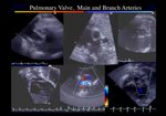
• The left upper pulmonary vein drains anomalously to a vertical vein, which drains into the innominate vein. Otherwise normal segmental cardiac anatomy. Normal biventricular size and systolic function.
MR:
• Impression: Normal pulmonary venous connections with partial anomalous drainage from the LUPV (white arrowhead) to a vertical vein (white arrow) to the in-nominate vein (yellow arrow). (Qp:QS 1.6 :1). Mildly dilated right ventricle with normal systolic function (EF 53%). Normal left ventricular size (LVEDV 69.53ml/m2) with normal systolic function (EF 55%). ↖ read less