Endoscopic Left L5-S1 Decompression in the Setting of a Prior L2-S1 Fusion
By
Usman Zahir
FEATURING
Usman Zahir
361 views
November 15, 2020
In this video a patient with a prior L2-S1 anterior/posterior fusion and right L5-S1 decompression ...
read more ↘ performed by another surgeon 6 years ago, underwent a left L5-S1 endoscopic decompression by me to resolve left leg pain from a large partially calcified disc herniation.
Most of my endoscopic cases are interlaminar (60-70%) with the rest transforaminal.
This is one such example of an interlaminar case. A 80+ year old male who came to me 6 years after a front back fusion L2-S1 and right L5-S1 hemilaminectomy. He presented with intractable left leg pain that he reports had been increasing since his operation ( he cant remember if the left leg pain was immediately after the operation or a few weeks/months later). Patient had been getting repeat steroid injections and his pain physician was running out of options.
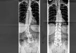
A few distracting pieces of information were a past CT scan from 3 years ago showing lucencies around S1 pedicle screws bilaterally suggesting pseudoarthrosis. However during my evaluation, he reports no back pain, just leg pain all in the S1 distribution on the left. There was no evidence of hardware impingement. Updated xrays this year also showed bridging bony fusion mass anteriorly at L5-S1 suggesting that he likely fused (just in a delayed fashion).
MRI performed this year showed a large left L5-S1 disc herniation partially calcified.
Its unclear how this disc herniation occurred in the setting of a L2-S1 fusion. I am suspecting that its possible disc material was iatrogenically displaced posteriorly when the ALIF cage was inserted. Another possibility is an incomplete discectomy during the ALIF, and that during the intervening months after surgery, since there was incomplete fusion, disc material herniated out the back due to motion at L5-S1.
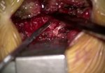
Nevertheless, this ended up being a fairly straightfrward case just with a lot of bony work. My plan was to do an indirect decompression of the disc but it ended up being less calcified than I expected. Just a lot of time was spent with the curved curette releasing the nerve from the disc.
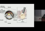
Endoscopic procedure was done through a small 1 centimeter incision. The prior screws and hardware were left in place.
↖ read less
Comments 0
Login to view comments.
Click here to Login


