Video Player is loading.
Current Time 0:00
/
Duration 0:00
Loaded: 0%
Stream Type LIVE
Remaining Time -0:00
1x
- 0.5x
- 0.75x
- 1x, selected
- 1.25x
- 1.5x
- 1.75x
- 2x
- Chapters
- descriptions off, selected
- captions settings, opens captions settings dialog
- captions off, selected
This is a modal window.
Beginning of dialog window. Escape will cancel and close the window.
End of dialog window.
10 seconds
Playback speed
This is a modal window. This modal can be closed by pressing the Escape key or activating the close button.
2,830 views
September 15, 2017
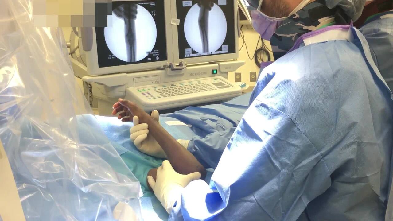
The purpose of this video is to demonstrate the surgical technique of closed reduction and ...
read more ↘ percutaneous pinning under fluoroscopic guidance of a displaced, pediatric extension type supracondylar humerus fracture. Intraoperative patient positioning, closed reduction maneuver, placement of 3 lateral smooth wires (0.062 inches) and postoperative splinting are demonstrated in this video. Pre-operative, intraoperative fluoroscopic and post-operative radiographic images are shown.
Title: Closed Reduction, Percutaneous Pinning of Supracondylar Humerus Fracture
Authors: Megan O’Connell, BS, David A. Fuller, MD, Cooper Medical School of Rowan University
Purpose: The purpose of this video is to demonstrate the surgical technique of closed reduction and percutaneous pinning under fluoroscopic guidance of a displaced, pediatric extension type supracondylar humerus fracture.
Methods: An 8 year old male with a displaced, extension type, extra-articular, supracondylar humerus fracture is shown undergoing closed reduction and percutaneous pinning of the fracture. Intraoperative patient positioning, closed reduction maneuver, placement of 3 lateral smooth wires (0.062 inches) and postoperative splinting are demonstrated in this video. Pre-operative, intraoperative fluoroscopic and post-operative radiographic images are shown.
Results: The video is 5min, 5 sec in duration.
Conclusion: Closed reduction and percutaneous pinning of a pediatric, supracondylar humerus fracture is demonstrated in this video.
↖ read less
Comments 3
Login to view comments.
Click here to Login