Playback speed
10 seconds
5,338 views
September 15, 2013
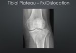
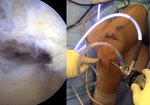
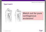
Arthroscopic fixation of Type III Meyers and McKeever displaced tibial eminence in a ten-years-old child. ...
read more ↘
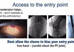
With the knee off the end of the table, a C-arm helps checking the tibial tunnels remain transepiphyseal, rather than transphyseal, to avoid physeal violation. A superomedial portal to have a good inflow of saline and two more ones, anterolateral viewing and anteromedial working suffice.
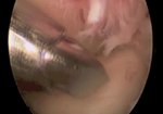
After debridement and fracture mobilization to evaluate reduction, the fragment is found completely dislodged from its origin, but reduction is easily achieved. No temporary fixation to secure the fragment.
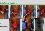
An ACL tibial guide was used to place two parallel drill tunnels by means of a 1.6 mm Kirschner wire, up and around the periphery of the anatomic footprint of the ACL and separated at least 1 centimeter apart at the outer epiphysis. The medial one is tangential to the eminence fragment and the lateral one is drilled through it.
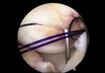
A pair of No. 0 braided silk sutures, atraumatically mounted into a straight needle, are passed backwards through each tibial tunnel up into the joint and through the tibial fragment. Both black silk threads are pulled loose with a suture retriever to develop two large loop that are left there for future use.
A hooked shuttling device is introduced, through the AM portal and the medial loop, to pierce the ACL in its mid-coronal plane and as close to the bony fragment as possible. A No. 1 PDS absorbable suture is shuttled into the knee and left there removing the hook out of the knee joint. The PDS suture is handled through the lateral loop with a suture retriever coming from the AM portal. The same sequence is repeated for the second PDS suture but the shuttling device pierces the ACL in its more anterior aspect.
The fracture is reduced by pulling down on the black silk suture ends, from both epiphyseal stab wounds. By careful suture management technique, each pair is separatedly passed to the epiphyseal stab wounds respectively and then tied with 4 or 5 knots over the bony bridge, while a gentle posterior drawer is applied.
A long leg cast is applied at 20 degrees of flexion.
↖ read less
Comments 6
Login to view comments.
Click here to Login


