Video Player is loading.
Current Time 0:00
/
Duration 0:00
Loaded: 0%
0:00
Stream Type LIVE
Remaining Time -0:00
1x
- 0.5x
- 0.75x
- 1x, selected
- 1.25x
- 1.5x
- 1.75x
- 2x
- Chapters
- descriptions off, selected
- captions settings, opens captions settings dialog
- captions off, selected
This is a modal window.
Beginning of dialog window. Escape will cancel and close the window.
End of dialog window.
10 seconds
Playback speed
This is a modal window. This modal can be closed by pressing the Escape key or activating the close button.
2,539 views
August 23, 2011
The deltoid and teres minor are critical for normal shoulder function and are innervated by ...
read more ↘ the axillary nerve. Patients with axillary nerve compression often describe posterior or lateral shoulder pain, weakness, and decreased athletic performance. In chronic cases, muscular atrophy may be present on examination. Magnetic resonance imaging can also be useful by revealing neurogenic edema or fatty infiltration of the deltoid or teres minor. It also provides the opportunity to evaluate soft tissue causes of compression and the presence of concomitant intraarticular shoulder pathology.
To this end, humeral osteophytes, malignancies, hypertrophied musculature, mal-united scapula fractures, and fibrous bands have each been identified as causes of axillary nerve compression.
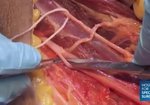
The axillary nerve travels just inferior to the subscapularis muscle belly and traverses inferior to the glenohumeral capsule to enter the quadrilateral space. It typically begins to arborizes near the long head of the triceps origin, forming several smaller branches. The more anterior branch wraps around the humeral neck and innervates the middle and anterior deltoid. The posterior branch is typically responsible for posterior deltoid, teres minor, and cutaneous innervation to the joint capsule and lateral shoulder.
The indications for axillary nerve decompression are not completely defined. The initial management is non-operative and patients are directed to discontinue aggravating activities and a general shoulder rehabilitation program prescribed. Failure to improve over a 3 to 6 month period, or the presence of large compressive lesions may indicate the need for surgical decompression.
Decompression of the axillary nerve using open techniques has been reported by a number of authors with satisfactory outcomes. A series by Cahill and Palmer reported 16 of 18 patients experienced dramatic or complete symptomatic relief after this procedure. McAdams and Dillingham reported on 4 overhead athletes with quadrilateral space syndrome who were able to return to full activities without pain at twelve weeks post-surgically. Smaller case reports have also documented successful surgical outcomes.
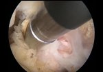
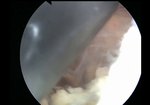
Arthroscopic transcapsular axillary nerve decompression is a novel procedure that we have primarily used to treat recalcitrant quadrilateral space syndrome and as part of our comprehensive arthroscopic management of glenohumeral degeneration in young patients with large impinging humeral osteophytes as a joint preservation procedure.
Though clinical studies comparing arthroscopic and open decompression techniques are not available it is reasonable to believe that an arthroscopic approach may facilitate visualization, and create less soft tissue disruption, thereby accelerating rehabilitation.
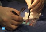
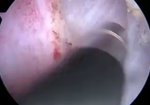
Therefore, we present our technique for arthroscopic, trans-capsular axillary nerve decompression in this video. When performed by experienced shoulder surgeons, it provides a relatively facile method to reliably decompress the axillary nerve.
↖ read less
Comments 1
Login to view comments.
Click here to Login

