Video Player is loading.
Current Time 0:00
/
Duration 0:00
Loaded: 0%
Stream Type LIVE
1x
- 0.5x
- 0.75x
- 1x, selected
- 1.25x
- 1.5x
- 1.75x
- 2x
- Chapters
- descriptions off, selected
- captions settings, opens captions settings dialog
- captions off, selected
This is a modal window.
Beginning of dialog window. Escape will cancel and close the window.
End of dialog window.
10 seconds
Playback speed
This is a modal window. This modal can be closed by pressing the Escape key or activating the close button.
Arthroscopic Evaluation of Lateral Ligaments of the Knee
685 views
October 30, 2019
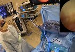
In this video I show how the lateral ligaments of the knee look like during ...
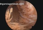
read more ↘ arthroscopy. You can see them from within the joint, comparing the healthy appearance of non injured knees with the pathologic looking in a knee with posterolateral corner injury.
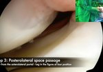
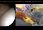
I explain the arthroscopic technique and leg position required to reach and see the lateral ligaments of the Knee.
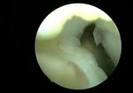
It includes the lateral collateral ligament ( or fibulo femoral ligament), anterolateral ligament, popliteus tendón, popliteus fibular ligament, hiatus popliteus.
The main challenge is to reach the hiatus popliteus region, what requires full extension of the knee.
In order to compare and help in identifying the injuries, I first show uninjured ligaments in healthy knees and later the torn ligaments in a pathologic knee.
Three patiens participate in this video. The first two had not injury in the lateral ligaments and the evaluation was made during knee arthroscopy for other reasons.
The third patient had a posterolateral corner injury with capsule torn, femoral dettachment of lateral colateral and anterolateral ligaments and partial injury in popliteus tendon. The popliteus-fibular ligament is seen through the posterolateral capsule defect.
↖ read less
Comments 1
Login to view comments.
Click here to Login


