Video Player is loading.
Current Time 0:00
/
Duration 0:00
Loaded: 0%
0:00
Stream Type LIVE
1x
- 0.5x
- 0.75x
- 1x, selected
- 1.25x
- 1.5x
- 1.75x
- 2x
- Chapters
- descriptions off, selected
- captions settings, opens captions settings dialog
- captions off, selected
This is a modal window.
Beginning of dialog window. Escape will cancel and close the window.
End of dialog window.
10 seconds
Playback speed
This is a modal window. This modal can be closed by pressing the Escape key or activating the close button.
Anterior Interosseous to Ulnar Motor Supercharge Nerve Transfer - Standard
4,188 views
April 10, 2012
<!--Code is buggy. I cannot use <br> or </br> and instead using </strong> for line ...
read more ↘ breaks-->
<strong>High Definition Standard Version - https://vimeo.com/40027121
High Definition Extended Version - https://vimeo.com/40108442
</strong>
<strong>Education Website - http://nervesurgery.wustl.edu
Video Library - https://vimeo.com/user6062882
</strong>
<strong>Anterior Interosseous to Ulnar Motor Supercharge Nerve Transfer</strong>
Standard Version (9.120404.120306)
</strong>
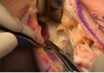
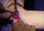
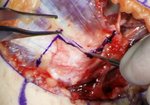
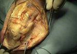
The supercharge nerve transfer is a procedure that coapts the distal end of a donor nerve to the side of the recipient nerve. Nerve regeneration is facilitated from donor to recipient through a perineurial window to enhance regeneration from the proximal regenerating nerve. This procedure can be used in cases of 2nd/3rd degree nerve injuries to augment motor recovery, with the advantages of a nerve transfer and without sacrificing the integrity and proximal regeneration of the recipient nerve. In this case, the patient had an iatrogenic medial cord injury. She presented 7 months following injury with ulnar intrinsic atrophy and fibrillations/motor unit potentials in her ulnar extrinsic/intrinsic muscles. The supercharge anterior interosseous to ulnar motor nerve transfer was elected with a transmuscular ulnar nerve transposition, Guyon’s canal release, and FDP tenodesis. This video highlights details of the supercharge nerve transfer, with specifics on the perineurial coaptation, Guyon’s canal release, and FDP tenodesis.
</strong>
Table of Contents
00:10 Orientation
00:15 Incision and Exposure
00:51 Identifying the Ulnar Nerve and Dorsal Cutaneous Nerve Branch
01:31 <strong>Guyon’s Canal Release</strong>
01:54 Identifying and Releasing the Palmar Carpal Ligament
02:07 Identifying and Releasing the Hypothenar Muscles
03:11 Exposing the Pronator Quadratus
03:22 Partial Release of the Flexor Digitorum Profundus Muscle Attachment
03:39 Identifying the Anterior Interosseous Nerve
03:50 Dividing the Pronator Quadratus
04:25 Transecting the Donor Anterior Interosseous Nerve
05:02 Neurolysis of the Ulnar Nerve and Creation of the Motor Perineurial Window
05:52 Anterior Interosseous to Ulnar Motor Supercharge Nerve Transfer
06:15 <strong>Flexor Digitorum Profundus Tenodesis</strong>
08:10 Scope Supercharge End-to-side Perineurial Window Repair
</strong>
Narration: Susan E. Mackinnon
Videography: Andrew Yee
</strong>
Terms of Use and Private Policy: nervesurgery.wustl.edu/pages/termsofuse.aspx
↖ read less
Comments 2
Login to view comments.
Click here to Login
