Video Player is loading.
Current Time 0:00
/
Duration 0:00
Loaded: 0%
0:00
Stream Type LIVE
Remaining Time -0:00
1x
- 0.5x
- 0.75x
- 1x, selected
- 1.25x
- 1.5x
- 1.75x
- 2x
- Chapters
- descriptions off, selected
- captions settings, opens captions settings dialog
- captions off, selected
This is a modal window.
Beginning of dialog window. Escape will cancel and close the window.
End of dialog window.
10 seconds
Playback speed
This is a modal window. This modal can be closed by pressing the Escape key or activating the close button.
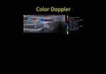
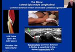
6 Sonographic Features of Tennis Elbow
By
Learn MSK Sono
FEATURING
Jamie Bie, RMSKS, RVT, RDMS
By
Learn MSK Sono
FEATURING
Jamie Bie, RMSKS, RVT, RDMS
141 views
June 12, 2023
Here are 6 sonographic features that represent the classic ultrasound findings associated with tennis elbow: ...
read more ↘
🎾 Osteophytes on the lateral epicondyle
🎾 Diffuse or focal hypoechoic, heterogeneous, and/or thickening of the common extensor tendon indicative of tendinopathy
🎾 Focal hypoechoic or anechoic disruption of tendon fibers due to varying degrees of tearing
🎾 Calcifications present within the common extensor tendon
🎾 Hyperemia present with the use of power doppler
🎾 Secondary thickening of the underlying Radial Collateral Ligament
↖ read less
Comments 0
Login to view comments.
Click here to Login