NYU Langone Orthopedics
- 2024 Comprehensive Spine Course
- Sixth International NYU Langone Hip Dysplasia Symposium
- 2022 Comprehensive Spine Course
- Fifth International NYU Langone Hip Dysplasia Symposium: The Island of Knowledge in Hip Dysplasia
- 2nd Annual Sports Medicine Symposium: The Golf and Soccer Athlete
- 2023 Featured Videos
- 2021 Fourth International Hip Dysplasia Symposium
- Featured Videos from NYU Langone Orthopedics
- 2021 Comprehensive Spine Course
- 2020 Comprehensive Spine Course
- 2019 Featured Videos from NYU Langone Orthopedics
- Sports Medicine Symposium: The Modern Athlete
- NYU Langone Orthopedic Webinar Series
- NYU Langone Orthopedics Faculty on VuMedi
- 44th Annual Howard Rosen Memorial Tri-State Trauma Symposium
- Events
Playback speed
10 seconds
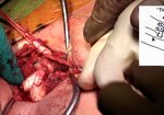
Osteochondral Allograft for a Full Thickness Articular Cartilage Defect of the Knee
3,586 views
August 22, 2012
Summary
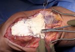
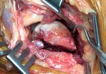
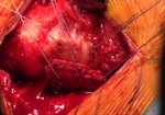
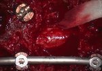
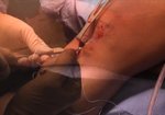
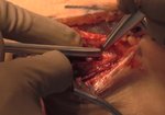
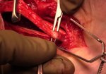
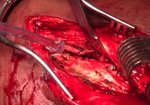
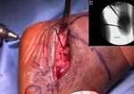
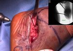
This video demonstrates an osteochondral allograft transplantation using the press-fit technique for a full ...
read more ↘ thickness articular cartilage defect of the knee.
Introduction
Chondral lesions of the knee are common, found in roughly 63% of knee arthroscopies in a study by Curl et al. Debridement, microfracture and osteochondral autograft transfer (OAT) have not been shown to be definitive in the treatment of articular cartilage lesions. Osteochondral allograft transplantation has shown encouraging results for large focal defects in younger patients and is a suitable treatment option.
Materials and Methods
Allograft preservation is an integral component of the treatment. Chondrocyte viability has been shown to decrease with time and must be stored at 4C. Studies have shown that by 3 weeks rougly 70% of chondrocytes are still viable while at seven weeks it is roughly 67% (Pearsall et al, 2004). Current guidelines recommend using the allograft before 42 days.
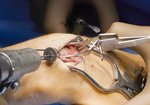
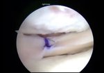
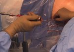
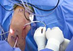
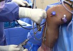
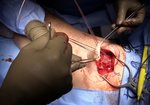
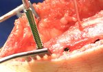
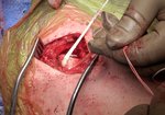
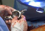
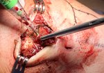
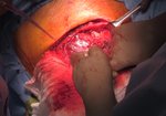
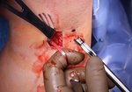
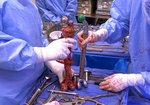
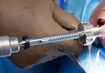
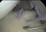
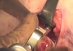
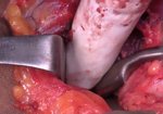
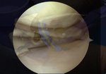
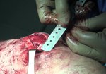
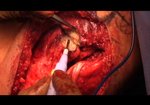
This video demonstrates the press-fit plug technique, whereby a guide wire is placed perpendicular to the articular surface. The lesion is then reamed to an appropriate size to a depth of 10 to 13 mm thickness. The depth of the recipient site is then measured at the 12 o’clock, 8 o’clock and 4 o’clock positions and the graft is sized accordingly on the ACT™ Allograft Implant Work Station and Vice (MTF, Inc, Edison, New Jersey). PRP is applied to the recipient site and the graft is gently tapped in taking care to match the clock position on the graft with the corresponding position at the recipient site.
Results
The literature has so far reported good clinical results using this technique with a graft survival rate ranging from 79%-100% at 3-10 years of follow-up (Demange and Gomoll, 2012). We report a case of a successful clinical outcome after osteochondral allograft transplantation for a full-thickness articular cartilage defect of the lateral femoral condyle.
Discussion
Press-fit technique for osteochondral allograft transplantation is a safe and effective treatment for large chondral and osteochondral lesions.
↖ read less
Comments 1
Login to view comments.
Click here to Login


